Introduction: What is homology?
Homology describes the biological features from a common evolutionary ancestor. [1] In genetics, two genes are homologous if they are derived from the same gene in a common ancestor. [2] Homologous genes are further separated into orthologs and paralogs. Orthologs are genes separated by speciation (different organisms), whereas paralogs are separated by gene duplication (same organisms).

Fig.1 Difference between homologs, paralogs and orthologs. [3]
Homologs of the HTRA1 gene
Model organisms that share the HTRA1 homologs. The information is gathered from BLAST, National Center for Biotechnology Information. <Click on each picture to access to FASTA>

|

|

|

|

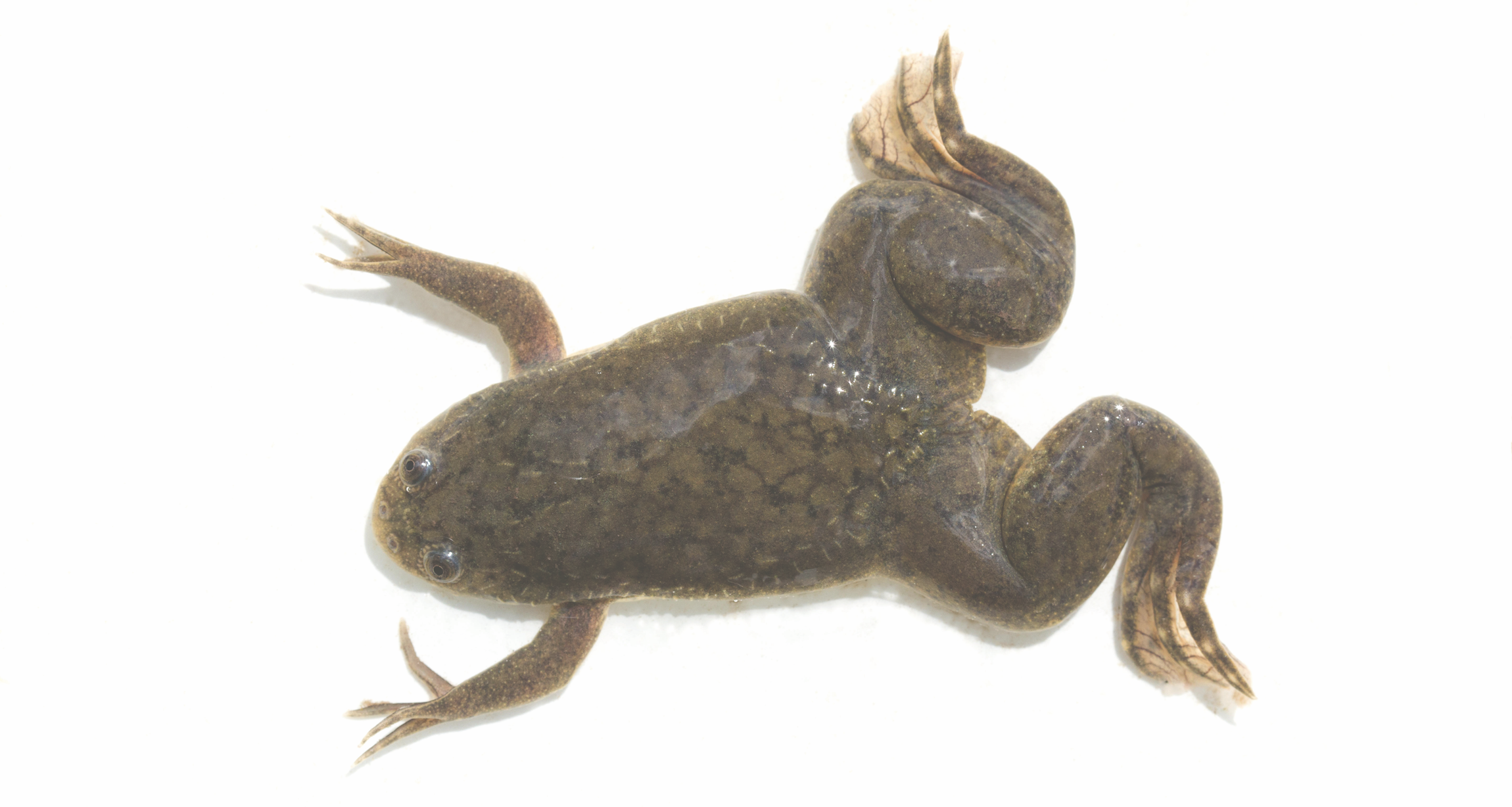
Western clawed frog (Xenopus tropicalis)
Accession code: XP_002932829.1 Length: 469aa Identity: 69.94% |
|

|

|
Choice of model organism
There is no best animal model that recapitulates all aspects of AMD, as AMD is a complex disease involving various genetic, environmental factors. Each model organism has its advantage and disadvantage in reflecting different aspects of AMD such as oxidative stress, lipid metabolism and inflammation, etc. Therefore, it is important to have a combination of different animal models for the study of AMD.
Mouse models: Based on the HTRA1 protein domain analysis and homolog comparison, the mouse model shares the most similarilies with human. Mice also have RPE, BrM, and choriocapillaris that are affected in AMD in human. However, mice do not have macular, and their retina is rod-dens
Non human primates(NHP) are the only pre-clinical animals that have a macula and share many similarities in eye structure organization. Some studies have demonstrated macaques to have early-onset drusen phenotype of AMD. However, they are expensive, difficult for genetically manipulation and require extensively long time study.[4]
Zebrafish models:
In my study, I decided to use zebrafish as the model organism for the following reasons.
Other in Vitro Models can also be used to study AMD
Primary RPE cells isolated from human fetal eyes (hfRPE), Stem Cell Derived RPE Cells (iPSC-RPE)[2]
Mouse models: Based on the HTRA1 protein domain analysis and homolog comparison, the mouse model shares the most similarilies with human. Mice also have RPE, BrM, and choriocapillaris that are affected in AMD in human. However, mice do not have macular, and their retina is rod-dens
Non human primates(NHP) are the only pre-clinical animals that have a macula and share many similarities in eye structure organization. Some studies have demonstrated macaques to have early-onset drusen phenotype of AMD. However, they are expensive, difficult for genetically manipulation and require extensively long time study.[4]
Zebrafish models:
In my study, I decided to use zebrafish as the model organism for the following reasons.
- Zebrafish requires lower cost and high reproduction rates.
- Visualization of blood vessel in zebrafish is good for angiogenesis study.
- Anatomically, zebrafish retina is cone-rich, which is similar to the human retina. This presents an advantage of zebrafish model for the study of AMD and other photoreceptor diseases over rodent models with rod-rich retina. Human macular is localized in retina and has highest concentration of cone photoreceptors across the retina. It is responsible for central, high-resolution, and color vision. [4] [6]
- Reliable model of neovascularization is able to be induced in zebrafish by putting them in hypoxia. [5]
- Around 70% of genes in the human genome have orthologs in the zebrafish genome. [5]
Other in Vitro Models can also be used to study AMD
Primary RPE cells isolated from human fetal eyes (hfRPE), Stem Cell Derived RPE Cells (iPSC-RPE)[2]
Discussion
From the FASTA data collected from NCBI using BLAST, we can see that HTRA1 is conserved across different species.
The conservation (%ID) decreases as the evolutionary distance increases. It is intriguing to note that HTRA1 is not found in yeast nor bacteria (E. coli), which limits the potential model organisms for the study of HTRA1 related disease. This indicates the unique role in more complicated organisms and inspires further investigations on the evolutionary development of HTRA1 gene.
The conservation (%ID) decreases as the evolutionary distance increases. It is intriguing to note that HTRA1 is not found in yeast nor bacteria (E. coli), which limits the potential model organisms for the study of HTRA1 related disease. This indicates the unique role in more complicated organisms and inspires further investigations on the evolutionary development of HTRA1 gene.
Reference:
human image :https://clipart-library.com/clip-art/human-silhouette-images-25.htm
Header:https://www.examplesof.net/2013/05/example-of-homologous-organs.html
[1]Quick guide. (n.d.). Retrieved from https://www.cell.com/current-biology/pdf/S0960-9822(04)00287-8.pdf
[2]Wagner, G. P. (2007). The developmental genetics of homology. Nature Reviews Genetics, 8(6), 473–479. https://doi.org/10.1038/nrg2099
[3]Bio, B. (2016, July 9). Homology Terminology: Never Say the Wrong Word Again. Retrieved March 29, 2024, from Bitesize Bio website: https://bitesizebio.com/26762/homology-terminology-never-say-wrong-word/
[4]Rho, J., Percelay, P., Pilkinton, S., Hollingsworth, T. J., Kornblau, I., & Jablonski, M. (2022). An Overview of Age-Related Macular Degeneration: Clinical, Pre-Clinical Animal Models and Bidirectional Translation. IntechOpen EBooks. https://doi.org/10.5772/intechopen.96601
[5]Rastoin, O., Gilles Pagès, & Maeva Dufies. (2020). Experimental Models in Neovascular Age Related Macular Degeneration. International Journal of Molecular Sciences (Online), 21(13), 4627–4627. https://doi.org/10.3390/ijms21134627
[6]Noel, N. C. L., MacDonald, I. M., & W Ted Allison. (2021). Zebrafish Models of Photoreceptor Dysfunction and Degeneration. Biomolecules, 11(1), 78–78. https://doi.org/10.3390/biom11010078
