Welcome!
DO YOU KNOW?
|
By 2030, 1 in
people in the world will be aged 60 years or over. [1]
|
An ageing world is posing new challenges to our health and societal systems, so are age-related diseases. AMD is one of them.
|
It is estimated that by 2040,
Million
people worldwide will have AMD. [2] |
What is Age-related Macular Degeneration?
Age-related macular degeneration (AMD) is an age-related eye disease that causes damage to macular, a yellow spot in the center of the retina. It is the leading cause of vision loss for over 60-year-old populations.
|
|
|
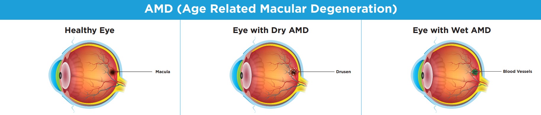
What happens to the eyes of AMD patients?
There are two types of AMD:
- Dry AMD (nonneovascular): The most common (80-90%) form of AMD that is accompanied by the accumulation of small yellow deposits called drusen between the retinal pigment epithelium (RPE) and Bruch’s membrane on the retina. [3]
- Wet AMD (neovascular, or nAMD): This form is less common (10~15%) but can cause more severe symptoms. Wet AMD is accompanied by the abnormal growth of blood vessels under the retina. These abnormal blood vessels tend to break and cause the leak of blood and other fluids, which causes the scarring of the macular. [4]
Anatomy of human eye
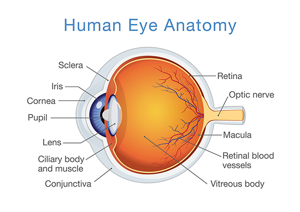
Human eye anatomy [5]
|
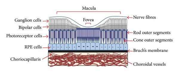
Diagram of macular layer structure [6]
|
What are the symptoms of AMD?
|

|
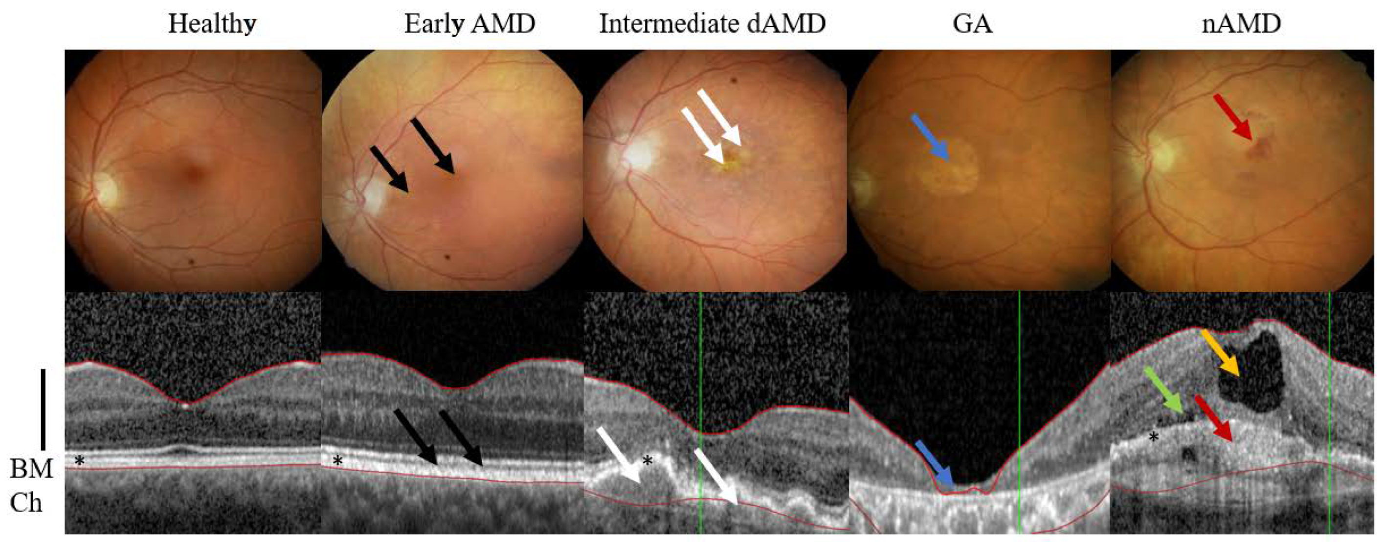
Different stage development of AMD [10]
|
The progression of AMD also involve several stages and there are many different ways to stratify it. According to the classification of Age-Related Eye Disease Study (AREDS), the stages of AMD can be as follows:
no AMD (no or few small drusen)
Geographic atrophy (GA) is an advanced form of dry AMD. It is characterized by atrophic lesion that progressively expand to cover the macular. As mentioned above, two types of AMD are not independent from each other, thus both GA and nAMD can coexist in one eye. [9] |
Diagnosis & Treatment
Diagonistic tools will be applied to definitively diagnose AMD including:
- A dialation test with eye drop.
- Optical coherence tomography (OCT), a non-invasive X-ray imaging method to take cross-section pictures of your retina to identify the thickening or thinninng of retina layers.
- Indocyanine green angiography (ICGA), which involves the injection of colored dye into your bloodstream and check for changes on your retina. [11]
|
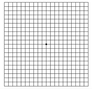
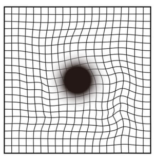
You can use the Amsler grid to monitor your vision. Cover each eye at one time and look directly at the dot in the center. If you see any parts bent, wavy or dim (as shown on the right), please contact your ophthalmologist for further examination. [12]
|
|
|
Currently there is no cure for AMD, and the treatments vary depending on the type of AMD. Here are treatments that have been reported to reduce AMD progressing:
Dry type treatments:
https://www.intechopen.com/chapters/75476
Wet type (nAMD) treatments:
There is also active research in developing gene therapies and stem cell therapies to treat vision loss caused by AMD, for more information, please click the corresponding button:
Dry type treatments:
- Although there is currently no proven treatment for dry AMD, potential therapeutic strategies have been proposed. For more information, please visit the literature:
https://www.intechopen.com/chapters/75476
Wet type (nAMD) treatments:
- Multiple injection of medicine monthly
- Nutritional suppliments with antioxidants
- Anti-VEGF treatments (intravitreal anti-vascular endothelial growth factor therapies)
There is also active research in developing gene therapies and stem cell therapies to treat vision loss caused by AMD, for more information, please click the corresponding button:
What are the risk factors of AMD?
|

|
How is HTRA1 gene related to AMD?
Human HTRA1 is located at 10q26.13. HTRA1 gene encodes high-temperature requirement protein A1 (HtrA1).This protein is a serine protease secreted by a number of tissues including retinal pigment epithelium (RPE).[13] This protein belongs to HtrA family and is evolutionary conserved, distributed from bacteria to humans. [14] The domain Increased expression of HTRA1 is identified to contribute to AMD in genomewide association studies (GWAS). [15][16]
However, HTRA1 is not the only gene that is associated with AMD. Other genetic factors, such as C3, CFH and CFHR3, are also reported by GWAS study to increase the risk of late-stage AMD. [17] Overall, AMD is a heterogeneous disease that involves genetics, metabolic and environmental factors. Physiological process of AMD involves oxidative stress, inflammation, dysregulated antioxidants, lipid metabolism, and angiogenesis. [18]
However, HTRA1 is not the only gene that is associated with AMD. Other genetic factors, such as C3, CFH and CFHR3, are also reported by GWAS study to increase the risk of late-stage AMD. [17] Overall, AMD is a heterogeneous disease that involves genetics, metabolic and environmental factors. Physiological process of AMD involves oxidative stress, inflammation, dysregulated antioxidants, lipid metabolism, and angiogenesis. [18]
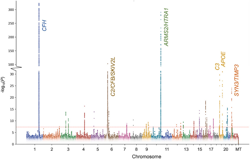
Manhattan plot of gene loci associated with AMD. Extracted from (Pan et al., 2023).
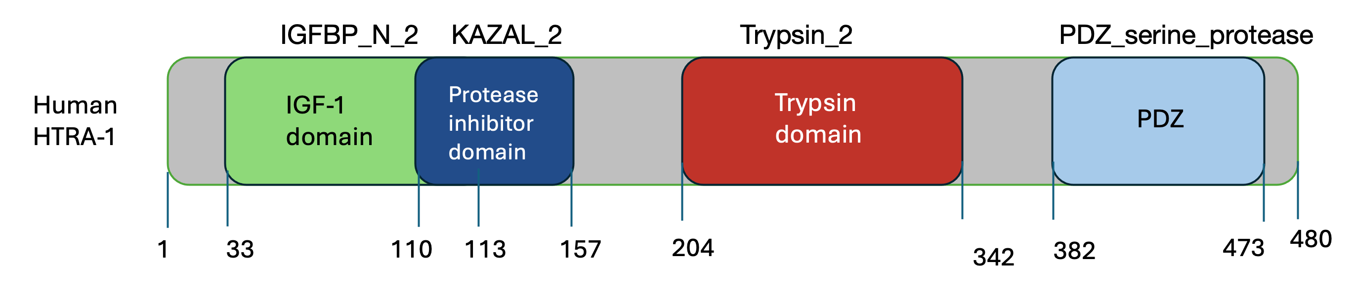
Domain organization of human HTRA-1 protein created by MS powerpoint
What is the gap in knowledge surrounding HTRA1?
The mechanism of how HTRA1 causes AMD still remains unknown. A recent study has demonstrated a long-range allosteric lock mechanism to inhibit the activity of HTRA1 to treat AMD.[18] However, an opposite role of HTRA1 that requires an enhancement in certain cancers and Alzheimer's disease [19] requires a further investigation of the side effects of inhibiting HTRA1 expression in treating AMD.
Further Information

|

|
Reference:
[1]Ageing and Health(2022). Retrieved from: https://www.who.int/news-room/fact-sheets/detail/ageing-and-health
[2]Wan Ling Wong, Su, X., Li, X., Cheung, M. G., Klein, R., Cheng, C.-Y., & Tien Yin Wong. (2014). Global prevalence of age-related macular degeneration and disease burden projection for 2020 and 2040: a systematic review and meta-analysis. The Lancet Global Health, 2(2), e106–e116. https://doi.org/10.1016/s2214-109x(13)70145-1
[3]Age-Related Macular Degeneration (AMD). Retrieved from: www.nei.nih.gov/learn-about-eye-health/eye-conditions-and-diseases/age-related-macular-degeneration
[4]Age-Related Macular Degeneration. Retrieved from: https://www.aao.org/eye-health/diseases/amd-macular-degeneration
[5]Human Eye Anatomy - Ophthalmologists and Retinal Specialists. (2022, July 12). Retrieved April 1, 2024, from Elman Retina Group website: https://www.elmanretina.com/services/human-eye-anatomy/
[6]Othman Al Gwairi, Thach, L., Wen Hua Zheng, & Little, P. J. (2016). Cellular and Molecular Pathology of Age-Related Macular Degeneration: Potential Role for Proteoglycans. Retrieved April 1, 2024, from ResearchGate website: https://www.researchgate.net/publication/305783621_Cellular_and_Molecular_Pathology_of_Age-Related_Macular_Degeneration_Potential_Role_for_Proteoglycans
[7]Sharon P., & Valerie K,.(2024) Age-related macular degeneration (AMD), Retrieved from: https://www.allaboutvision.com/conditions/amd.htm
[8]Age-Related Macular Degeneration: Current Treatments and Future Therapies. Retrieved from: https://www.youtube.com/watch?v=ftZfPYgMkkc&ab_channel=StanfordHealthCare
[8]Rho, J., Percelay, P., Pilkinton, S., Hollingsworth, T. J., Kornblau, I., & Jablonski, M. (2022). An Overview of Age-Related Macular Degeneration: Clinical, Pre-Clinical Animal Models and Bidirectional Translation. IntechOpen EBooks. https://doi.org/10.5772/intechopen.96601
[9]Bakri, S. J., Bektas, M., Sharp, D., Luo, R., Sarda, S. P., & Khan, S. (2023). Geographic atrophy: Mechanism of disease, pathophysiology, and role of the complement system. Journal of Managed Care & Specialty Pharmacy, 29(5-a Suppl), S2–S11. https://doi.org/10.18553/jmcp.2023.29.5-a.s2
[10]Heloterä, H., & Kaarniranta, K. (2022). A Linkage between Angiogenesis and Inflammation in Neovascular Age-Related Macular Degeneration. Cells, 11(21), 3453–3453. https://doi.org/10.3390/cells11213453
[11]Dry macular degeneration. Retrived from:https://www.mayoclinic.org/diseases-conditions/dry-macular-degeneration/symptoms-causes/syc-20350375
[12]Gheorghe, A., Mahdi, L., & Musat, O. (2015). Age-Related Macular Degeneration. Romanian journal of ophthalmology, 59(2), 74–77.
[13]Gerhardy, S., Ultsch, M., Tang, W. et al. Allosteric inhibition of HTRA1 activity by a conformational lock mechanism to treat age-related macular degeneration. Nat Commun 13, 5222 (2022). https://doi.org/10.1038/s41467-022-32760-9
[14]Chien, J. et al. A candidate tumor suppressor HtrA1 is downregulated in ovarian cancer. Oncogene 23, 1636–44. (2004).
[15] Fritsche LG, Fariss RN, Stambolian D, Abecasis GR, Curcio CA, Swaroop A. Age-related macular degeneration: genetics and biology coming together. Annu Rev Genomics Hum Genet. 2014;15:151-71. doi: 10.1146/annurev-
[16]Lin MK, Yang J, Hsu CW, Gore A, Bassuk AG, Brown LM, et al. HTRA1, an age-related macular degeneration protease, processes extracellular matrix proteins EFEMP1 and TSP1. Aging Cell 2018;17:e12710.
[17] Borchert, G. A., Hoda Shamsnajafabadi, Hu, M. L., De, S. R., Downes, S. M., MacLaren, R. E., … Jasmina Cehajic-Kapetanovic. (2023). The Role of Inflammation in Age-Related Macular Degeneration—Therapeutic Landscapes in Geographic Atrophy. Cells, 12(16), 2092–2092. https://doi.org/10.3390/cells12162092
[18]Lin MK, Yang J, Hsu CW, Gore A, Bassuk AG, Brown LM, et al. HTRA1, an age-related macular degeneration protease, processes extracellular matrix proteins EFEMP1 and TSP1. Aging Cell 2018;17:e12710.
[19]Poepsel, S. et al. Determinants of amyloid fibril degradation by the PDZ protease HTRA1. Nat. Chem. Biol. 11, 862–869 (2015).
Images:
https://unsplash.com/s/photos/genetics
https://www.fightingblindness.ca/eyehealth/eye-diseases/age-related-macular-degeneration/
https://allaboutvision.com/conditions/amd.htm
https://lhblind.org/what-is-age-related-macular-degeneration-amd/
|
|
|
